Compact Bone Diagram Lacunae : Compact Bone Spongy Bone And Other Bone Components Human Anatomy And Physiology Lab Bsb 141 : To know the architecture of compact and spongy (cancellous) bone.
Compact Bone Diagram Lacunae : Compact Bone Spongy Bone And Other Bone Components Human Anatomy And Physiology Lab Bsb 141 : To know the architecture of compact and spongy (cancellous) bone.. Compact bone diagram bone cross section diagram file624 diagram of compact bone new. Compact bone makes up 80 percent of the human skeleton. Rather, the osteocytes containing lacunae are arranged in a. These structures are brought into motion by skeletal muscles. The osteocytes are sitting in the lacunae and the canals are canaliculi, which interconnect the lacunae with the major vessels.
The three dimensional functional units within compact bone are called osteons. A structural unit of compact bone consisting central haversian canal. To know the architecture of compact and spongy (cancellous) bone. It can be remodeled all throughout life to withstand stress. These structures are brought into motion by skeletal muscles.
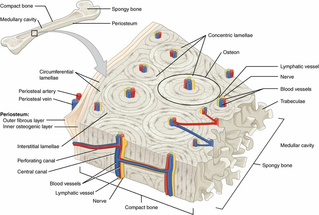
A P Bone Unit 4 Flashcards Quizlet from o.quizlet.com Compact bone diagram osteon compact bone ap pinterest anatomy human anatomy and. Compact bone , also called cortical bone , dense bone in which the bony matrix is solidly filled with organic ground substance and inorganic salts, leaving only tiny spaces (lacunae) that contain the osteocytes , or bone cells. Lacunae, small chambers containing osteocytes, are arranged concentrically around the central canal. Between the rings of the matrix, the bone cells (osteocytes) are small channels (canaliculi) radiate from the lacunae to the osteonic (haversian) canal to provide. A diagram of the anatomy of a bone, showing the compact bone. The osteon consists of a central canal called the osteonic (haversian) canal, which is surrounded by concentric rings (lamellae) of matrix. 6 compact bone vs spongy bone. Like compact bone, spongy bone, also known as cancellous bone, contains osteocytes housed in lacunae, but they are not arranged in concentric circles.
Compact bone is sometimes called cortical bone.
Once the osteoid is mineralized, the precursor cells get surrounded by organic intracellular substances called lacunae to become fully developed and matured into osteocytes. To know the structures of a synovial joint and a symphysis joint (intervertebral disc). This shows the architecture of compact bone which is designed to nourish and regulate osteocytes and bone matrix. In histology, a lacuna is a small space, containing an osteocyte in bone, or chondrocyte in cartilage. 31.01.2021 · compact bone, also called cortical bone, dense bone in which the bony matrix is solidly filled with organic ground substance and inorganic salts, leaving only tiny spaces (lacunae) that contain the osteocytes, or related posts of compact bone diagram labeled skeleton bones diagram. 6 compact bone vs spongy bone. The walls of the diaphysis are composed of dense and hard compact bone. Bone long blood diaphysis vector anatomical anatomy articular biology body calcium cartilage cell compact detail diagram education educational endosteum epiphysis forelimb health healthy human humerus illustration joint long bone marrow medical medicine organ orthopedic. Although compact bone is made up of haversian systems, it is almost solid. Compact and cancellous — or spongy osteocytes, or bone cells, are found in lacunae, which are spaces within the lamellae. Like compact bone, spongy bone, also known as cancellous bone, contains osteocytes housed in lacunae, but they are not arranged in concentric circles. Interstitial lamellae are located between osteons. Between the rings of the matrix, the bone cells (osteocytes) are small channels (canaliculi) radiate from the lacunae to the osteonic (haversian) canal to provide.
Start studying compact bone structure. Like compact bone, spongy bone, also known as cancellous bone, contains osteocytes housed in lacunae, but they are not arranged in concentric circles. The three dimensional functional units within compact bone are called osteons. Pig bone diagram wiring diagram, femur bone diagram full human skeleton diagram femur simple anatomy, colored ear diagram for kids bone labeled of the eye to label compact bone diagram simple diagram system. A diagram of the anatomy of a bone, showing the compact bone.
Compact Bone Diagram Anatomy And Physiology Human Anatomy And Physiology Physiology from i.pinimg.com A structural unit of compact bone consisting central haversian canal. The walls of the diaphysis are composed of dense and hard compact bone. You should include the histology of compact bone slides with diagram as well into your article. Compact bone consists of closely packed osteons or haversian systems. Basically, in kindergarten when you drew skeletons, you were drawing compact bone. Compact bone surrounds the spongy bone tissue and it has a unique appearance. Compact bone is sometimes called cortical bone. This makes it very dense.
It can be remodeled all throughout life to withstand stress.
Basically, in kindergarten when you drew skeletons, you were drawing compact bone. Between the rings of the matrix, the bone cells (osteocytes) are small channels (canaliculi) radiate from the lacunae to the osteonic (haversian) canal to provide. You should include the histology of compact bone slides with diagram as well into your article. Compact bone consists of closely packed osteons or haversian systems. Explore more like lacunae in compact bone. This makes it very dense. Lesion composed of dense cortical bone (compact bone) with definite osteocyte lacunae and cement lines (line visible by microscopic examination marking the boundary of an osteon/ haversian system). Lacunae, small chambers containing osteocytes, are arranged concentrically around the central canal. These structures are brought into motion by skeletal muscles. Lacunae are the small spaces in bone tissue where mature bone cells called osteocytes are. A structural unit of compact bone consisting central haversian canal. Compact bone diagram osteon compact bone ap pinterest anatomy human anatomy and. Like compact bone, spongy bone, also known as cancellous bone, contains osteocytes housed in lacunae, but they are not arranged in concentric circles.
The osteon consists of a central canal called the osteonic (haversian) canal, which is surrounded by concentric rings (lamellae) of matrix. Learn vocabulary, terms and more with flashcards, games and other study tools. To know the architecture of compact and spongy (cancellous) bone. Although compact bone is made up of haversian systems, it is almost solid. Compact and cancellous — or spongy osteocytes, or bone cells, are found in lacunae, which are spaces within the lamellae.
Compact Bone Structure Biology Dictionary from biologydictionary.net A diagram of the anatomy of a bone, showing the compact bone. This makes it very dense. Lesion composed of dense cortical bone (compact bone) with definite osteocyte lacunae and cement lines (line visible by microscopic examination marking the boundary of an osteon/ haversian system). Compact bone surrounds the spongy bone tissue and it has a unique appearance. Compact bone consists of closely packed osteons or haversian systems. Once the osteoid is mineralized, the precursor cells get surrounded by organic intracellular substances called lacunae to become fully developed and matured into osteocytes. Compact bone diagram bone cross section diagram file624 diagram of compact bone new. In an ordinary microscopic section, viewed by transmitted light, they appear as fusiform opaque spots.
Compact bone consists of closely packed osteons or haversian systems.
You should include the histology of compact bone slides with diagram as well into your article. Bone osseous tissue labeled cancellous bone structure spongy bone diagram compact bone connective tissue vascular lacunae compact bone 400x haversian system bone osteocyte function osteoblast osteocyte osteoclast bone cell. Lacunae are the small spaces in bone tissue where mature bone cells called osteocytes are. The basic units of compact bone are called osteons or haversian systems. The lacunae are situated between the lamellae, and consist of a number of oblong spaces. To know the structures of a synovial joint and a symphysis joint (intervertebral disc). A diagram of the anatomy of a bone, showing the compact bone. The musculoskeletal system is comprised of bones and connective tissue structures, such as cartilage, ligaments, and tendons. It can be remodeled all throughout life to withstand stress. Compact bone makes up 80 percent of the human skeleton. Compact bone , also called cortical bone , dense bone in which the bony matrix is solidly filled with organic ground substance and inorganic salts, leaving only tiny spaces (lacunae) that contain the osteocytes , or bone cells. Compact bone surrounds the spongy bone tissue and it has a unique appearance. Structure of bone diagram 9 photos of the structure of bone diagram bone cell diagram, bone composition, bone marrow diagram, bone structure and function, bone structure of the human body, bone structure worksheet, compact bone diagram, types of joints, human anatomy.
Learn vocabulary, terms and more with flashcards, games and other study tools compact bone diagram. You should include the histology of compact bone slides with diagram as well into your article.


0 Komentar