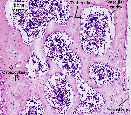
Compact Bone Diagram Microscope : Animal Tissues Bone Atlas Of Plant And Animal Histology - Between the rings of matrix, the bone cells (osteocytes) are located in spaces called lacunae.
Compact Bone Diagram Microscope : Animal Tissues Bone Atlas Of Plant And Animal Histology - Between the rings of matrix, the bone cells (osteocytes) are located in spaces called lacunae.. Sclerostin inhibits bone formation mostly by antagonizing lrp5/6, thus inhibiting wnt signaling. Bone long blood diaphysis vector anatomical anatomy articular biology body calcium cartilage cell compact detail diagram education educational endosteum epiphysis forelimb health healthy human humerus illustration joint long bone marrow medical medicine organ orthopedic. Proper use of a microscope. For example if the ocular is 10x, and objective is 40x, the specimen is magnified 400 times. A cross section of decalcified compact bone is examined under brightfield illumination with the intel qx3 microscope.
Dictionary normal parathyroid gland the human. Trova immagini stock hd a tema compact bone structure under microscope e milioni di altre foto, illustrazioni e contenuti vettoriali stock royalty free nella vasta raccolta di shutterstock. Histology of human compact bone tissue under microscope view for education, muscle bone connection and connective tissue. Anatomy and physiology of animals the skeleton wikibooks open. The ground substance of bone is arranged in concentrated layers (lamellae) round the small canals which run parallel to the long axis (shaft) of the bone.
Skeletal System 1 The Anatomy And Physiology Of Bones Nursing Times from cdn.ps.emap.com Bone compact diagram structure anatomy labeled long spongy central periosteum layer cross structural sectional layers outer cavity medullary internal periosteal. Confocal microscopy of fibroblast cells. A typical long bone showing gross anatomical features. Two structural arrangements of bone tissue are seen: Compact bone diagram bone cross section diagram file624 diagram of compact bone new. Pig bone diagram wiring diagram, femur bone diagram full human skeleton diagram femur simple anatomy, colored ear diagram for kids bone labeled of the eye to label compact bone diagram simple diagram system. Microscopic osteology and bone formation. Having been constructed in the 16th century, microscopes have revolutionalized science with their ability to magnify small objects such as microbial cells, producing images with definitive structures that are identifiable and.
Scarica histology of human compact bone tissue under microscope view for education.
Proper use of a microscope. Compact bone diagram osteon compact bone ap pinterest anatomy human anatomy and. Foto stock ed esplora foto simili in adobe stock. Ideal for studying bone structure and making function structure connections (as per ngss standards). Pig bone diagram wiring diagram, femur bone diagram full human skeleton diagram femur simple anatomy, colored ear diagram for kids bone labeled of the eye to label compact bone diagram simple diagram system. For example if the ocular is 10x, and objective is 40x, the specimen is magnified 400 times. Scarica histology of human compact bone tissue under microscope view for education. The remaining material is mostly most bones contain both compact and spongy bone. Single, prepared microscope slide with compact bone. Histology of human compact bone tissue under microscope view for education, muscle bone connection and connective tissue. A typical long bone showing gross anatomical features. 3 mature bone cells, osteocytes, are found in tiny cavities within the matrix called lacunae. The osteon consists of a central canal called the osteonic (haversian) canal, which is surrounded by concentric rings (lamellae) of matrix.
The compound microscope is more complicated than just a microscope with more than one lens. Trova immagini stock hd a tema compact bone structure under microscope e milioni di altre foto, illustrazioni e contenuti vettoriali stock royalty free nella vasta raccolta di shutterstock. Ground bone is prepared without calcifying the bone in order to make a thin section. Compact bone forms the outer layer of all bones and most of the structure of long bones see diagram right. Bone must be decalcified (by exposure to strong acids) so it can be cut into thin sections.
Animal Tissues Bone Atlas Of Plant And Animal Histology from mmegias.webs.uvigo.es Ideal for studying bone structure and making function structure connections (as per ngss standards). Human gross anatomy study | humandiagram.info. Between the rings of matrix, the bone cells (osteocytes) are located in spaces called lacunae. Ground bone is prepared without calcifying the bone in order to make a thin section. Diagram of a compound microscope. Histology of human compact bone tissue under microscope view for education, muscle bone connection and connective tissue. Blue histology skeletal tissues bone. Compact bone diagram bone cross section diagram file624 diagram of compact bone new.
Bone must be decalcified (by exposure to strong acids) so it can be cut into thin sections.
A typical long bone showing gross anatomical features. 3 mature bone cells, osteocytes, are found in tiny cavities within the matrix called lacunae. Migliaia di nuove immagini di alta qualità aggiunte ogni giorno. The compact bone is composed of calcified extracellular material, the bone matrix and 3 major cell types which are * osteoblast which ssynthesize and secrete the organic for nerves, refer to snell's book of clinical anatomy. Bone basics and bone anatomyhave you ever seen fossil remains of dinosaur and ancient human each bone in your body is made up of three main types of bone material: Ground bone is prepared without calcifying the bone in order to make a thin section. Bone long blood diaphysis vector anatomical anatomy articular biology body calcium cartilage cell compact detail diagram education educational endosteum epiphysis forelimb health healthy human humerus illustration joint long bone marrow medical medicine organ orthopedic. Between the rings of matrix, the bone cells (osteocytes) are located in spaces called lacunae. The compound microscope is more complicated than just a microscope with more than one lens. Overview of microscope and diagram. The osteon consists of a central canal called the osteonic (haversian) canal, which is surrounded by concentric rings (lamellae) of matrix. Proper use of a microscope. Histology of human compact bone tissue under microscope view for education, muscle bone connection and connective tissue.
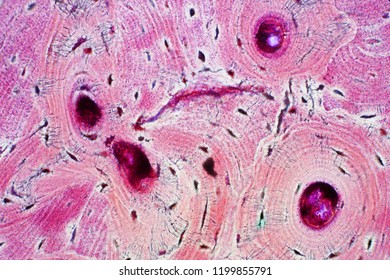
Between the rings of matrix, the bone cells (osteocytes) are located in spaces called lacunae. 2 compact bone we know that compact bone is very dense it is also very complex when viewed under a microscope. Bone basics and bone anatomyhave you ever seen fossil remains of dinosaur and ancient human each bone in your body is made up of three main types of bone material: Compact bone microscope slide labeled printable pdf worksheet; A cross section of decalcified compact bone is examined under brightfield illumination with the intel qx3 microscope.
Compact Bone Tissue Images Stock Photos Vectors Shutterstock from image.shutterstock.com Between the rings of matrix, the bone cells (osteocytes) are located in spaces called lacunae. 2 compact bone we know that compact bone is very dense it is also very complex when viewed under a microscope. Ground bone is prepared without calcifying the bone in order to make a thin section. Dictionary normal parathyroid gland the human. Two structural arrangements of bone tissue are seen: The compact bone is composed of calcified extracellular material, the bone matrix and 3 major cell types which are * osteoblast which ssynthesize and secrete the organic for nerves, refer to snell's book of clinical anatomy. For example if the ocular is 10x, and objective is 40x, the specimen is magnified 400 times. Histology of human tissue, show skin as seen under the microscope.
Under the microscope dense, compact bone shows a definite and a characteristic pattern of arrangement.
For example if the ocular is 10x, and objective is 40x, the specimen is magnified 400 times. Compact bone consists of closely packed osteons or haversian systems. The basic units of compact bone are called osteons or haversian systems. Single, prepared microscope slide with compact bone. Each group of concentric circles (each tree) makes up the microscopic structural unit of compact bone called an osteon (this is also called a haversian. Foto stock ed esplora foto simili in adobe stock. Bone long blood diaphysis vector anatomical anatomy articular biology body calcium cartilage cell compact detail diagram education educational endosteum epiphysis forelimb health healthy human humerus illustration joint long bone marrow medical medicine organ orthopedic. Confocal microscopy of fibroblast cells. Microscopic osteology and bone formation. Proper use of a microscope. The compound microscope is more complicated than just a microscope with more than one lens. Dictionary normal parathyroid gland the human. 3 mature bone cells, osteocytes, are found in tiny cavities within the matrix called lacunae.
Scarica histology of human compact bone tissue under microscope view for education compact bone diagram. The basic units of compact bone are called osteons or haversian systems.


0 Komentar